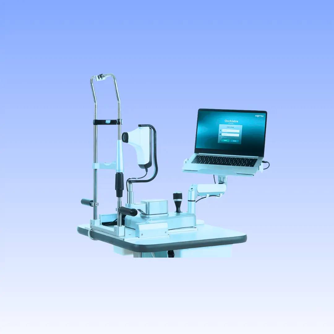
Main Features
Auto meibography
- Evaluate the meibomian glands with red light, the software provides automatic evaluation of loss area.
Non-invasive breakup time
- Automatically analyze break-up area, first and average break-up time for tear stability evaluation.
Interferometry
- Record a video of blinking process to observe the surface reflection pattern and dynamics of the tear film.
Tear meniscus height
- Automatically evaluate tear meniscus height that is observed on the eyelid margins. Up to 5 measurement points can be taken.
Fluorescein Staining
- Evaluate the areas of damage on the ocular surface after application of the fluorescein dye. Compare your images with grading scales incorporated in the software.
Auto Redness
- Eye redness could be one of the symptoms of dry eye disease. Automatically compare your images with grading scales incorporated in the software.
Eyelid margin imaging
- MGD can cause the glands to become blocked, impacted, and infected. Capture high resolution under white LED illumination, and compare your images with grading scales included in the software .
Color-coded reports for quick dry eye insights
- The DEA Dry Eye Analyzer's easy-to-read report template with color coding transforms data interpretation into an intuitive experience.
Specifications
IMAGE AND VIDEO ACQUISITION
Image Resolution 8,000,000 pixels
Image Dimensions 3864 x 2218 pixels JPEG
Acquisition Mode Multi-shot photos, video
Focus and Exposure Manual and automatic
Covering Area Maximum 8 mm
Camera Colored, sensitive to infrared
Light Source Red, blue and white LED
GENERAL INFORMATION
Working Distance 5 mm – 50 mm
Ports USB 3.0
Power Supply 5 V
Dimensiones 170 mm (F) × 54 mm (W) × 64–110 mm (L)
Weight 427 g (including main body, 4 lenses and 1 wireless camera shutter)
Accessories Standard: wireless camera shutter, lenses, briefcase, lens case slit lamp adaptor
Optional: complete holder, instrument
table
SOFTWARE AND DATA MANAGEMENT
Operating System Windows 10 64 bits
System Requirement Intel Core i3, RAM 8 GB, hard disk 200 GB,
screen resolution: 1920*1080
Exams DEQ-5, fluorescein staining, NIBUT, FBUT, interferometry, auto meibomian gland
evaluation, auto tear meniscus height, auto redness, eyelid margin imaging, anterior imaging